

Best Scalp Camera for Salon Use: What to Compare First
The best scalp camera for salon use is the one that gives clear, repeatable images. Compare magnification, lighting, storage, display and tip hygiene.
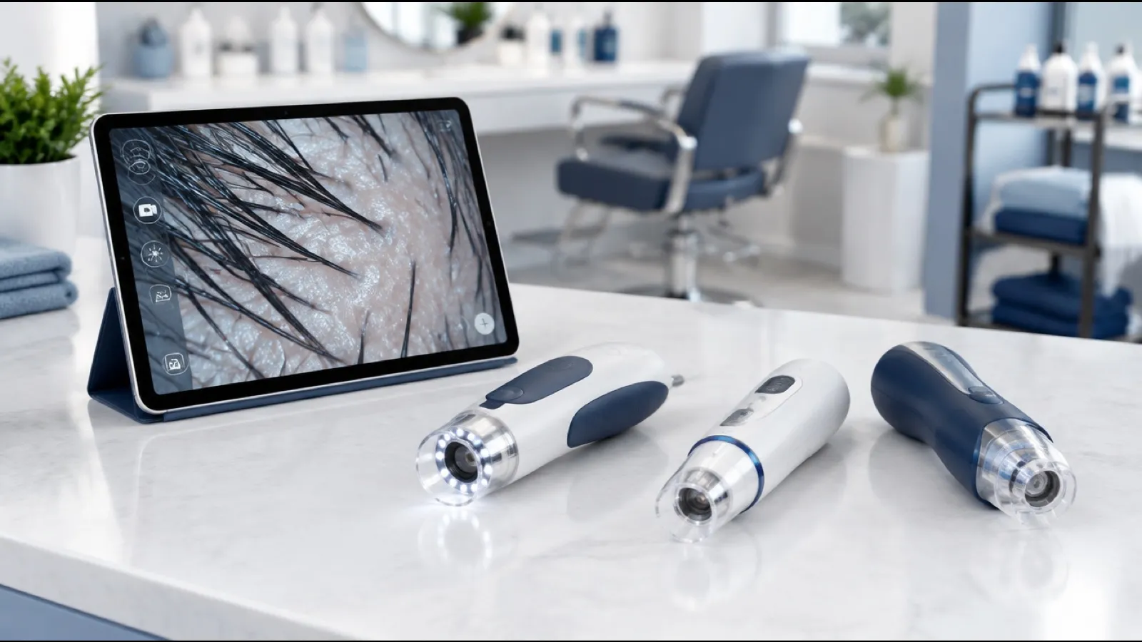
The best scalp camera for salon use is the one that gives you clear, evenly lit, repeatable images of the same scalp zones at every visit. Judge it on six features: magnification range, lighting, connection, software and image storage, a client-facing display, and tips you can keep hygienic. Brand and the biggest magnification number matter far less.
This guide compares features, not brands. It does not rank products or quote specifications for any model, because sellers change them and many are not independently verified. Use the criteria below to test whatever device you are considering.
What is the best scalp camera for a salon?
The best scalp camera for a salon is a device that shows both a wide view of the scalp surface and a close view of follicle openings, lights the image evenly, saves photos to a client record, and can be cleaned between clients. If two devices do those things, choose the one your team will use every day.
Sellers use several names for much the same tool: scalp camera, scalp scope, scalp microscope, scalp analyzer, trichoscope. A "scalp analyzer" usually means a camera bundled with software that labels or scores the image. Treat any automated score as a talking point for the consultation. It is not a finding, and it is never a diagnosis.
What features should you compare on a scalp scope?
Compare six things on every scalp scope: the magnification range, whether it has polarized and non-polarized light, how it connects, what the software does with images, whether the client can see the screen, and how the tips are cleaned. Ask the seller for unedited sample images at each setting before you pay.
| Feature | Why it matters | What to check before buying |
|---|---|---|
| Magnification range | Different structures need different views. See the next table. | Sample images at the lowest and highest setting. Ask how the stated number was measured, since the size of an image on screen also depends on the display. |
| Polarized vs non-polarized light | Polarized light cuts surface glare. Non-polarized light shows the surface as it reflects. | Which modes the device has, and whether you can switch during a session. |
| Wired vs wireless | Wired devices take power and data through a cable. Wireless devices move freely but rely on battery and connection. | Compatibility with your phone, tablet or computer. Battery life across a full day. What happens when the connection drops. |
| Software and image storage | The value of a scalp camera is the record it builds over time. | Whether images save with date, client name and zone. Where files are stored, how they export, who can open them, and whether there is an ongoing fee. |
| Client-facing display | Clients believe what they can see. | Whether the image appears on a screen visible from the chair, and whether you can show two dates side by side. |
| Hygiene of tips | The tip touches or comes close to the scalp. | Whether tips are removable, what they are made of, whether spares are sold, and the maker's written cleaning and disinfection instructions. |
What magnification do you need for each structure?
You need at least two working views. A paper in the International Journal of Trichology states that a manual dermoscope gives about 10x magnification and that videodermoscopes use lenses ranging from 20x to 1000x. That is the range used in medicine. A salon does not need all of it.
| What you want to see | View needed | What a good image shows |
|---|---|---|
| Scalp surface | Lower end of the device's range, wide field | A section of the part line, skin color, flaking, oil, buildup |
| Follicle openings | Middle of the range | Individual openings and how many hairs come out of each |
| Hair shaft | Higher end of the range | Single hairs in focus, so thickness can be compared between neighbors |
If a device cannot hold focus at its highest setting in your hand, that setting is not useful in a salon. Test it on a real scalp, not a printed card.
Polarized or non-polarized light?
Both have a use. The same paper notes that the patterns between follicles are visualized only with a polarizing light source or filter, and that blood vessel patterns are best seen without direct contact, because pressure on the skin can blanch them. Those are findings from medical practice. The practical point for a salon is simpler: a device with both modes shows you more, and you should use one consistent mode for comparison photos.
Wired or wireless?
Choose on reliability in your own space. A wireless scope is easy to move around the chair and pass over the head. A wired scope avoids battery and pairing problems. Either one fails as a consultation tool if it takes several minutes to connect while a client waits.
Trichoscope vs dermatoscope: what is the difference?
A dermatoscope is a handheld magnifier with built-in light that physicians use to examine skin. Trichoscopy is the use of dermoscopic imaging on the hair and scalp. A trichoscope is a dermatoscope or video device sold for that purpose. The hardware overlaps. The difference that matters is who is using it and why.
The International Journal of Trichology paper cited above is titled "Trichoscopy in Alopecias: Diagnosis Simplified", and it describes trichoscopy as a method for diagnosing types of hair loss. It lists signs that physicians are trained to interpret, such as yellow dots, black dots and variation in hair shaft diameter. As a diagnostic method, trichoscopy is a physician's tool.
A trichoscope for hairstylists is the same kind of camera used for a different job: to observe, document and decide whether to refer. Trichologists are not physicians either, and "trichologist" is not a state-licensed medical title in the US. For how the two roles differ, see trichologist vs dermatologist.
What can and can't you conclude from scalp camera images?
You can conclude what is visible. You cannot conclude what is causing it. A scalp microscope shows surface detail that the naked eye misses, but an image alone does not give a non-medical professional the history, examination, lab work or biopsy that a physician may use to reach a diagnosis.
| You can say | You cannot say |
|---|---|
| "There is flaking around the follicle openings at the crown." | "You have seborrheic dermatitis." |
| "These hairs look thinner than the ones next to them." | "This is genetic hair loss." |
| "Compared with your photo from the last visit, this area looks the same." | "Your hair is growing back." |
| "I can't see follicle openings in this patch. A dermatologist should look at it." | "These follicles are dead." |
| "There is product residue on the scalp surface." | "This buildup is causing your hair loss." |
The left column describes. The right column diagnoses or predicts. Stay on the left, and write your notes the same way.
How do you read scalp scope images as a beginner?
Start by looking at the same four things in every image: the scalp surface, the follicle openings, the hair shafts, and anything that differs from the surrounding area. Record each as a plain observation. Learning how to read scalp scope images means learning to describe consistently before you try to interpret.
A beginner's observation list:
- Skin color. Even tone, pink or red areas, or darker patches. Note where.
- Scale and flaking. Fine and dry, or thicker and oily looking. White or yellowish. Loose on the surface or attached around openings.
- Oil and residue. Shine on the skin, or film and particles that look like product.
- Follicle openings. Visible or not visible. Evenly spaced or with gaps.
- Hairs per opening. One, two, or more hairs emerging from each opening.
- Hair thickness. Similar from hair to hair, or a mix of thick and fine hairs in the same field.
- Short or broken hairs. Hairs much shorter than the rest, and whether the ends look tapered or snapped.
- Dots at the openings. Any dark or yellowish dots where a hair would be. Record the color and location only.
- Client response. Tenderness, itch or pain when the tip touches the area.
Do not attach condition names to any of these. The step-by-step scan, zone map and photo routine are covered in our scalp analysis protocol for hairstylists. If you see smooth bare patches, shiny skin with no visible openings, pustules, crusting, or pain, follow the checklist in when to refer a client to a dermatologist.
How do you keep scalp camera tips hygienic?
Clean and disinfect any part that touches the scalp after every client, using a method that both your state board rules and the device maker allow. Removable tips are easier to manage than fixed ones. If the maker gives no written cleaning instructions, treat that as a reason not to buy.
A workable routine:
- Remove the tip after the scan.
- Clean off visible oil, scale and product.
- Disinfect with a product your state board accepts for non-porous implements, for the contact time on its label.
- Let it dry, then store it covered.
- Keep enough spare tips to rotate through a busy day.
Do not scan over broken skin, open sores or weeping areas. Stop, document what you saw with the naked eye, and refer.
How do you choose and start using one?
Shortlist two or three devices that pass the feature table, test each on several scalps, and pick the one that gives the clearest repeatable images with the least setup. Then practice on existing clients before you charge for the service. You can compare options on the USTI scalp scopes page.
The camera will show you more than cosmetology or barber school trained you to describe. USTI's scalp health courses and the trichology pathway teach what you are looking at on screen and where your role ends. Once you are confident with the tool, it can anchor a paid service. See how to add scalp treatments to a salon menu.
The best scalp camera for salon work is the one that ends up in your hand at every consultation, produces images you can compare over months, and keeps you describing what you see while a physician handles diagnosis.
New to the field? Start with What is trichology? or look up a term in the trichology glossary.
Sources
- Jain N, Doshi B, Khopkar U. Trichoscopy in Alopecias: Diagnosis Simplified. International Journal of Trichology, 2013. Full text read via Europe PMC.
- American Academy of Dermatology: Hair loss: Who gets and causes
- National Institute of Arthritis and Musculoskeletal and Skin Diseases: Alopecia areata
Frequently asked questions
What is the best scalp camera for a salon?
There is no single best model. The best scalp camera for a salon is the one that produces sharp, evenly lit images at a wide view and a close view, saves them with a date and client name, shows them on a screen the client can see, and has tips you can clean between clients. Compare devices on those features.
What magnification does a scalp scope need?
No standard sets a required number for salon use. Published trichoscopy literature describes handheld dermoscopes at about 10x and video devices with lenses from 20x to 1000x. For salon work you need a lower setting that shows a part line and scalp surface, and a higher setting that shows follicle openings and individual hair shafts.
What is the difference between a trichoscope and a dermatoscope?
A dermatoscope is a magnifying device with built-in light that physicians use to examine skin. Trichoscopy is the use of that kind of device on the hair and scalp. A trichoscope is a dermatoscope or video camera sold for hair and scalp use. The tools overlap. What differs is who uses them and for what purpose.
Can a hairstylist use a trichoscope?
A hairstylist can use a magnifying scalp camera to observe and photograph the scalp as part of a consultation. A hairstylist cannot use it to diagnose a hair or scalp condition. Diagnosis is a physician's job. Service rules vary by state, so check with your state board about what your license covers before you sell a scalp analysis.
Is polarized light better on a scalp camera?
It shows different things. A review in the International Journal of Trichology notes that patterns between the follicles are seen only with a polarizing light source or filter. Polarized light reduces surface glare. Non-polarized light shows the surface as it reflects. A device that offers both modes gives you more options. Keep the mode the same for follow-up photos.
Is a wireless scalp camera better than a wired one?
Neither is better in every salon. Wireless devices are easier to move around the chair but depend on battery life and a stable connection. Wired devices draw power from the phone, tablet or computer and tend to connect the same way each time. Test either type with the screen and software you already own before buying.
What can I tell a client from scalp scope images?
You can describe what is visible: flaking, redness, oil, buildup, how many hairs come from each opening, and whether hairs look similar in thickness. You cannot name a condition, state a cause, or predict regrowth. If the image shows something that looks medical, recommend a dermatologist and offer the client copies of the photos.
This article is educational and is not medical advice. USTI teaches professionals to recognize, support and refer.
Turn this knowledge into a certification.
Learn hair and scalp science with USTI educators, from your first course to clinical training.
Explore trichology coursesRelated articles
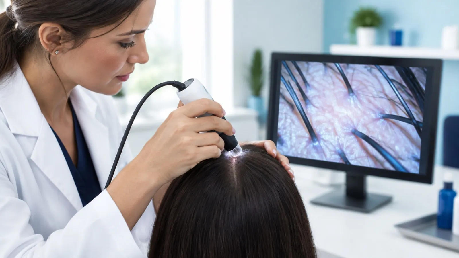
What Is Trichoscopy? Scalp Dermoscopy Explained
What is trichoscopy? A plain guide to scalp dermoscopy: how a trichoscope works, what it shows, who uses it, and how it compares with a biopsy or pull test.

Hair and Scalp Consultation: Full Guide and Intake Form
A hair and scalp consultation in six stages, with a ready-to-copy intake question list, an observation checklist, summary wording and a follow-up schedule.
How to Talk to a Client About Hair Loss: 6 Salon Scripts
How to talk to a client about hair loss: word-for-word scripts for six salon situations, a say-this-not-that table, privacy rules and referral wording.